| Microtome (Spencer Type) : |
|
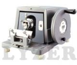
Lyzer Senior Rotary Microtome (LT-41) : (820 Spencer type) latest model, with one razor 120mm and honing stone. Feed range 150 micron in steps of one micron with automatic feed release device, complete in box with three holders, oil can and raxine cover. |
|
 |
|
| Microtome Semi Automatic: |
|
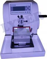
Lyzer Semi Automatic Microtome: Digital Control Panel Technical Specifications: 1. Motorized drive.
2. Compatible Embedding Material: Paraffin, Plastic.
3. Range of the thickness of slice: 0.25 - 60 μm.
Range of the thickness of trimming: 10 - 100 μm.
4. Setting thickness of slice ranges:
0.25----1 μm increment 0.25 μm
1----10 μm increment 1 μm
10----20 μm increment 2 μm
20----60 μm increment 5 μm.
5. Specimen Horizontal feeding: 28 mm.
6. Stroke length: 60 mm.
7. Knife index: High Profile Disposable Blades
8. Maximum section of slice: 50 x 50 mm.
9. Retractable specimen clamp, lateral knife holder 10. Weight:24 kg.
11. Working Voltage: 220 V, 50 Hz.(110 V, 60 Hz. optional)
12. Delivery time: 25 Days. |
|
 |
|
|
|
Mirror : College grade (concave or convex) highly silvered, optically true with beautiful ring on its outer surface, special quality, F.L.10, 20, 25cm
Diameter :
a) 50mm
b) 75mm |
|
 |
|
|
|
With gall bladder showing all structures and lobes clearly. All parts numbered and may be identified with the attached key card, mounted on stand.
Dimensions overall 200 X 90 X 250 mm. |
 |
|
|
|
Showing external details, large size mounted on stand.
Dimensions overall 170 X 90 X 21 mm. |
 |
|
|
|
Showing details of typical mesophytic leaf, mounted on board.
Dimensions overall 410 X 250 X 65 mm. |
 |
|
|
|
Showing complete internal details of the root of smilax, mounted on board. Dimensions overall 330 X 250 X 65 mm. |
 |
|
|
|
Showing various tessues vascular bundles in transverse section of a monocot stem. Sounted on board.
Dimensions overall 350 x 350 x 65mm. & asexual Clamydomonas L. H. |
 |
|
| MONOCOT SYTEMANTOMYT.S. & L.S.MAIZE |
|
This model exhibits the various tisses and the scattered closed and collateral vascular bundles in transverseand lonitudinal section. The large pitted vessels, spiral and annular vessels show the cell wall thickeninges in L.S. Very useful model for teaching the anatomy of monocot stem.
Mounted on base with key card 500 X 105 X 350 mm. |
 |
|
| Monocular Microscope (LT-10) |
|
Research Monocular Microscope (LT-10) : With quadruple revolving nose peice, detachable mechanical stage, 360 degree revolving Monocular head with hard coated prisms, graduated precise slow motion, with built in light arrangement having 6v-20w halogen lamp and solid state light control arrangement complete in thermocole box with following optics. Eye pieces : WF10 x and H 5x
Obejectives : 4x, 10x, 40x (SL), 100x (SL) oil |
|
 |
|


